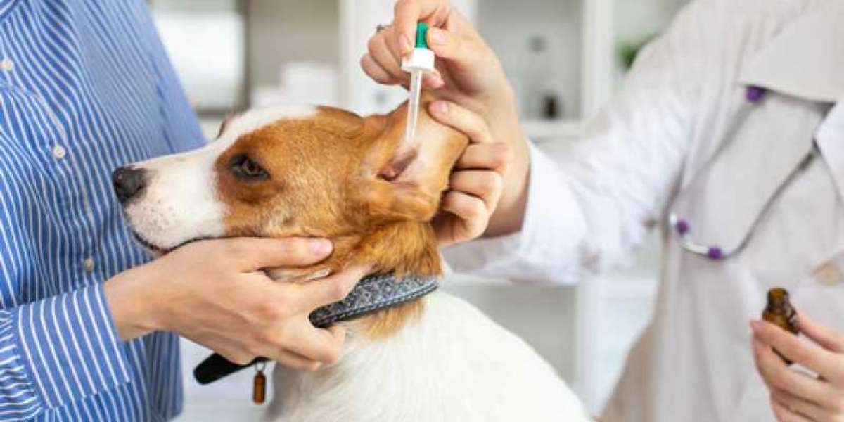
 Whether computed tomography, CT scan or common radiology x-rays, this technology is instrumental in diagnostics today. A CT scan can show gallbladder, kidney, or bladder stones, making it straightforward for the vet to determine the reason for illness. For instance it's important that bones heal correctly and in the proper course, meaning a visible picture can't be overstated. These images can inform the vet on the best plan of action, from surgery to letting nature take its course. As everyone knows, most dogs love to chew on stuff, and generally they swallow that stuff. A vet will often have an excellent hunch if the bone is damaged or not, but the x-ray will verify any suspicion. Many times that injury will be hidden beneath the skin, and may be too painful for the vet to palpate.
DoveLewis is the one facility in Oregon to be licensed on any degree (I, II or III). An echocardiogram can be a useful check accomplished at the aspect of an intensive bodily exam, a well-taken history of medical signs, and other exams like an ECG and chest x-rays. While many bigger veterinary hospitals have their own ultrasound gear, secondary practices don't. Once a prognosis, treatment, and follow-up plan are established, it is essential to comply with the treatment plan exactly and maintain routine follow-up evaluations with your veterinarian to ensure your canine has one of the best end result. If your canine does not require any sedation before their echocardiogram, they will eat and drink normally. By the time you see visible indicators of a coronary heart downside — difficulty respiration, rapid respiration, coughing, weak point, lethargy, exercise intolerance and collapsing — your canine might have coronary heart illness.
Your FAQs on Pet Echocardiograms Answered
Determining in case your dog has coronary heart illness, and what stage they’re in, allows your veterinarian to provide one of the best remedy plan. Purina’s new Pro Plan Veterinary Diet CardioCare is a prescription food plan that's backed by analysis that confirmed the food slowed the development of cardiac illness in its early levels. Hill’s Prescription Diet Heart Care h/d and Royal Canin Veterinary Diet Early Cardiac are different stable options. These diets are sodium-restricted, which helps prevent fluid accumulation and helps wholesome blood stress — both of that are necessary for cardiac patients. The nutrients include antioxidants, anti-inflammatories and different things that support cardiac perform. Cherished Companions Animal Clinic is a veterinary clinic in Castle Rock, Colorado.
Caring for Pets in Tracy
Final board certification requires profitable completion of a rigorous board examination. The cardiologists at MedVet are committed to continually bettering their knowledge and genoma laboratório Veterinário practice by pursuing continuing schooling even after passing their board examinations. They additionally present persevering with training to the veterinary group each regionally and nationally. Sometimes, a veterinary heart specialist or sonographer may recommend a chest X-ray to examine for symptoms associated to heart issues. For example, fluid in your cat or canine's lungs may level to congestive heart failure. These insights enable us to develop an individualized treatment plan to accurately handle and handle your cat's illness. If another checks need to be accomplished to assist diagnose your pet’s coronary heart situation, the heart specialist or technician will discuss this suggestion with you previous to performing these checks.
Why Would My Pet Need an Echocardiogram?
Doppler (both Color Doppler and Spectral Doppler), is another non-invasive ultrasound take a look at used to assess how blood is flowing through the center, in addition to how blood enters and exits it.
La medición de la agilidad de la sangre la empleamos en diversas patologías cardíacas, no solo para el diagnóstico, sino asimismo para el pronóstico y el tratamiento. Como ejemplo coloco un caso de degeneración valvular mitral en un perro que vino a la clínica presentando un soplo en el corazón. Como se puede observar en la imagen superior durante la sístole aparece un chorro de sangre que no va del ventrículo a la aorta, sino retorna a la aurícula izquierda, es lo que hace aparición en color. La ecocardiografía o ecografía del corazón es la técnica diagnóstica más ampliamente usada para la evaluación no invasiva de las patologías cardiovasculares. La ecocardiografía se volvió una técnica diagnóstica esencial en cardiología canina y felina, a tal punto, que sin su realización un diagnóstico cardiológico queda en el aire. En la ecocardiografía observamos la causa de dicho encharcamiento pulmonar, el líquido presiona al corazón, realizando que se colapse el atrio derecho, por lo que el ventrículo derecho recibe poca sangre para impulsarla a los pulmones.
Ecocardiografía
Aprovechamos también para analizar dicho líquido y poder comprender el origen del mismo y tratarlo. La gata del vídeo tiene por nombre Margarita, vino a la clínica en colapso al no llegarle oxígeno a los tejidos. Debimos ponerla en un ámbito abundante en oxígeno antes de empezar a explorarla, y después hacerlo por pasos, alternando con la cámara de oxígeno, ya que al cabo de unos minutos de estar fuera de la cámara se asfixiaba. Construcciones que sí se ven en la radiografía de la derecha, y que coloco para cotejar, es de un caso de asma felino, en el que al estar los pulmones hiperinflaccionados, el aire hace un mejor contraste. La relevancia de conocer el flujo y sentido de la sangre lo muestro con el caso de una gatita que íbamos a operar de castración y a la que le encontramos un soplo cardiaco en la exploración.


